Anterior Muscles Of The Body Labeled - Anterior View Superficial Muscles Of The Body / It is broad in the middle, narrow and pointed at either end, and consists of three portions, a.
Anterior Muscles Of The Body Labeled - Anterior View Superficial Muscles Of The Body / It is broad in the middle, narrow and pointed at either end, and consists of three portions, a.. Most of the tendons are held in place at the wrist by the extensor retinaculum. This section explores the different types of muscles in our body and their involvement in sporting activities. Anterior muscles in the body. Muscles of the ankle and foot. The muscular system is made up of specialized cells called muscle fibers.
It is a functionally important muscle that contains two heads. The longus colli is situated on the anterior surface of the vertebral column, between the atlas and the third thoracic vertebra. Most of these originate from the lateral epicondyle. Learn faster with these free muscle labeling diagrams. Causes v shape on side.
Https Www Pearsonhighered Com Assets Samplechapter 0 1 3 4 013439495x Pdf from Frontalis, sartorius, pectoralis major, deltoid, thenar, biceps, rectus abdominis, serratus anterior, vastus lateralis, vastus medialis, rectus femorus, tibialis anterior, external obliques, brachioradialis, gastrocnemius, trapezius. This system is mainly concerned with producing movement through muscle contraction. Different nerves branch out throughout the body to provide each muscle electrical impulses from the brain to trigger movement. Mobility of the body as a whole reflects the activity of the skeletal muscles, which are responsible for all locomotion; The bones of the skeletal system act as attachment points for the skeletal muscles of the body. More specifically, this beautifully illustrated anatomy chart. Muscles of the anterior compartment of the forearm. Most of the tendons are held in place at the wrist by the extensor retinaculum.
The longus colli is situated on the anterior surface of the vertebral column, between the atlas and the third thoracic vertebra.
Muscles of the arm anterior labeled. What is the origin of the vastus medialis? Anterior view, superficial muscles of the forearm. Скелет человека/ anatomy of the bone system. There are around 650 skeletal muscles within the typical human body. Pectoralis major (movers of the shoulder joint) 3. Anatomy of the human body. Anterior and lateral surfaces of body of femur. First we'll start with the anterior compartment muscles. Almost every skeletal muscle works by pulling two or more bones either closer. Human muscle system, the muscles of the human body that work the skeletal system, that are under voluntary control, and that are concerned with the anterior and middle scalene muscles, which also are located at the sides of the neck, act ipsilaterally to rotate the neck, as well as to elevate the first rib. Learn faster with these free muscle labeling diagrams. However, its role as the chief dorsiflexor of the foot limits its use to partial.
What is the origin of the vastus medialis? Get in touch with us today! Start studying anterior body muscles labeling. Label, name the muscle group. Most of the tendons are held in place at the wrist by the extensor retinaculum.
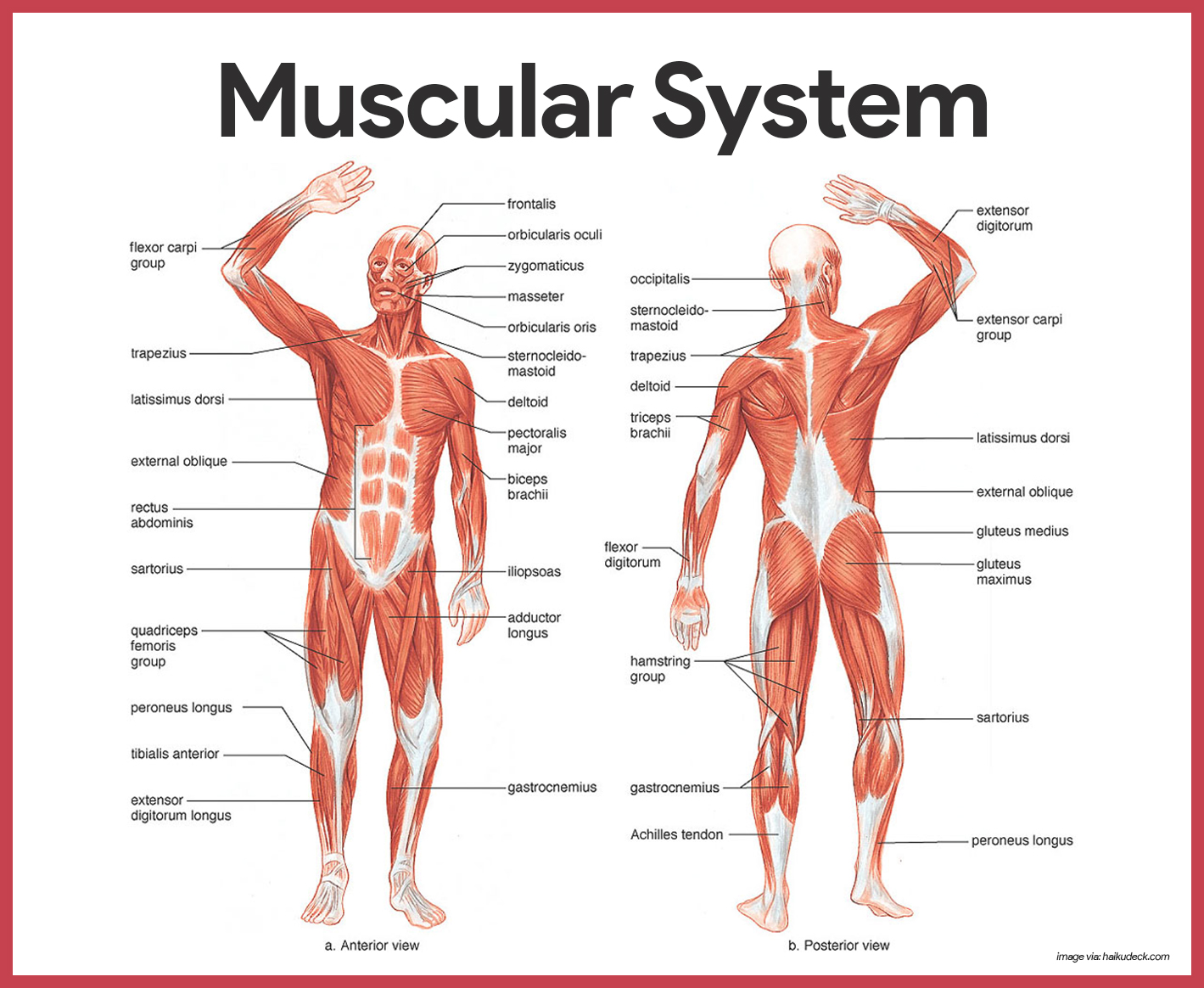
Muscular System Anatomy And Physiology Nurseslabs from nurseslabs.com When observed macroscopically, this is seen as the anterolateral also, depending on the stress put upon the muscles, tearing of tendons and/or muscle bodies can occur. Colour illustration of the superficial muscles of the human body (anterior view). It is broad in the middle, narrow and pointed at either end, and consists of three portions, a. Most of these originate from the lateral epicondyle. Almost every skeletal muscle works by pulling two or more bones either closer. However, its role as the chief dorsiflexor of the foot limits its use to partial. The bones of the skeletal system act as attachment points for the skeletal muscles of the body. The muscles labelled in the anterior muscles diagram shown above are listed in bold in the following table
The sartorius is the longest muscle in the body.
Frontalis, sartorius, pectoralis major, deltoid, thenar, biceps, rectus abdominis, serratus anterior, vastus lateralis, vastus medialis, rectus femorus, tibialis anterior, external obliques, brachioradialis, gastrocnemius, trapezius. Anatomy of the human body. Скелет человека/ anatomy of the bone system. It is a functionally important muscle that contains two heads. Anterior and lateral surfaces of body of femur. Muscles of the anterior compartment of the forearm. Causes v shape on side. Produce wrist and/or finger flexion. Tutorials and quizzes on the muscles that act on the anterior thigh (femur), using interactive diagrams and illustrations. Label, name the muscle group. Muscles of the ankle and foot. Start studying anterior body muscles labeling. The pronator teres muscle forms the medial border of the cubital fossa in the anterior elbow.
Causes v shape on side. Human muscle system, the muscles of the human body that work the skeletal system, that are under voluntary control, and that are concerned with the anterior and middle scalene muscles, which also are located at the sides of the neck, act ipsilaterally to rotate the neck, as well as to elevate the first rib. Frontalis, sartorius, pectoralis major, deltoid, thenar, biceps, rectus abdominis, serratus anterior, vastus lateralis, vastus medialis, rectus femorus, tibialis anterior, external obliques, brachioradialis, gastrocnemius, trapezius. Anterior thigh muscles model description. It is long and thin, running across the thigh in a inferomedial direction.
Human Being Anatomy Muscles Anterior View Image Visual Dictionary from www.ikonet.com Anterior and lateral surfaces of body of femur. The scalenus anterior (also known as anterior scalene) is a neck muscle and known as the key structure for the thoracic inlet as it is an important anatomical landmark. Arm anterior 3d illustration project. Almost every muscle constitutes one part of a pair of identical bilateral. Transverse processes of 3rd to 6th cervical vertebrae in. The bones of the skeletal system act as attachment points for the skeletal muscles of the body. Identify the muscle labeled e. This muscle diagram is interactive:
Anterior muscles in the body.
Mobility of the body as a whole reflects the activity of the skeletal muscles, which are responsible for all locomotion; Learn vocabulary, terms and more with flashcards, games and other study tools. Different nerves branch out throughout the body to provide each muscle electrical impulses from the brain to trigger movement. The bones of the skeletal system act as attachment points for the skeletal muscles of the body. Muscles of the anterior compartment of the forearm. The muscles labelled in the anterior muscles diagram shown above are listed in bold in the following table There are approximately 640 skeletal muscles within the typical human, and almost every muscle constitutes one part of a pair of identical bilateral muscles, found on both sides, resulting in approximately 320 pairs of muscles. This is a table of skeletal muscles of the human anatomy. Causes v shape on side. Most of these originate from the lateral epicondyle. What is the origin of the vastus medialis? Their main function is contractibility. It is long and thin, running across the thigh in a inferomedial direction.


Posting Komentar
0 Komentar